Treatment
2D and 3D CBCT imaging
Imaging should answer a question. This page sets out which questions each type of image answers, and why CBCT is not taken routinely.
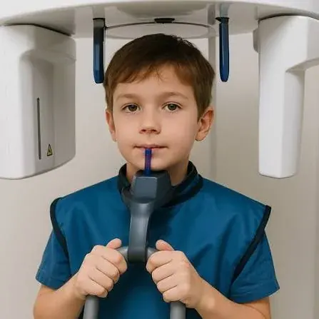
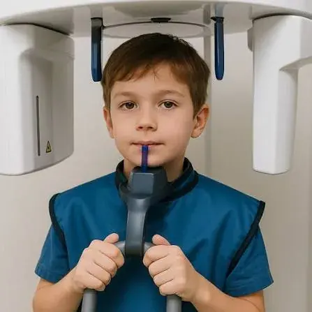
What each image shows
| Image | What it shows | Typical use |
|---|---|---|
| Panoramic (OPG) | A flat survey of both jaws, all teeth, developing teeth and gross pathology | Standard orthodontic screening; presence and position of unerupted teeth |
| Lateral cephalogram | A standardised side view of the skull and jaws | Skeletal relationships, incisor inclination, growth analysis and treatment planning |
| Periapical / occlusal | A detailed view of a small area | Root morphology, resorption, localisation of an impacted tooth |
| Cone beam CT (CBCT) | A three-dimensional volume of bone and teeth | Suture maturation before MARPE, impacted tooth localisation, root proximity, joint bone changes, miniscrew site planning |
When CBCT is justified
CBCT delivers more radiation than a panoramic radiograph, though less than medical CT. It is justified where a two-dimensional image cannot answer the clinical question and the answer will change what is done. In this practice the commonest genuine indications are:
- Assessment of mid-palatal suture maturation before bone-anchored expansion.
- Localisation of an impacted tooth and its relationship to adjacent roots, particularly upper canines.
- Planning miniscrew or bone plate sites where bone depth must be known.
- Assessment of condylar bone morphology where degenerative change or asymmetry is suspected.
- Assessment of alveolar bone before movements that risk fenestration or dehiscence.
Reporting
A three-dimensional volume contains structures well outside the orthodontic field of interest. Formal reporting means the whole volume is examined, so that incidental findings are identified and referred rather than missed.
Frequently asked questions
Is a CBCT scan safe for children?
The same principles apply, with more caution: children are more radiosensitive, so the justification threshold is higher and the field of view is kept as small as the clinical question allows. Most children need no CBCT at all.
Can I bring scans taken elsewhere?
Yes, and it is encouraged. Recent imaging from another clinic often avoids repeating an examination. Bring the original data file where possible, not just a printed report.
How often are radiographs repeated?
Only when a new clinical question arises — typically at diagnosis, sometimes during treatment where progress needs checking, and at the end where indicated. Not on a fixed schedule.
Page last reviewed: 2026-09-10. This page is general health information about orthodontic care and is not a diagnosis, a treatment plan, or a substitute for an in-person clinical examination. Suitability for any treatment described here can only be established after a consultation, clinical assessment and the appropriate records. If you have symptoms that concern you, seek advice from a qualified clinician.
Appointments
Arrange a specialist orthodontic assessment in Dubai
Consultations take place at Manhal Medical Center, Al Safa 1, Dubai, in English or Arabic. Every assessment begins with a full clinical examination and, where indicated, 3D imaging — not a treatment quote.