Treatment
Radiology reporting
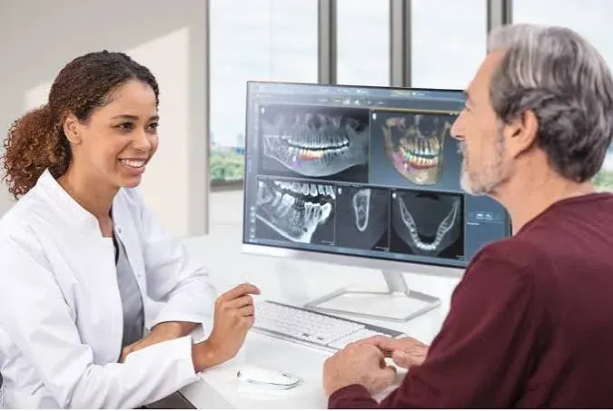
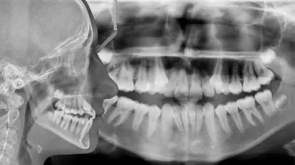
Taking an image and reading an image are different acts. Formal reporting means the whole volume is examined, not only the part the clinician was interested in.
Why it matters
A cone beam CT taken to assess a mid-palatal suture also contains the maxillary sinuses, the nasal cavity, the temporomandibular joints, the cervical spine within the field of view, and every tooth and its supporting bone. A clinician looking only at the suture will not see anything else — and the responsibility for what appears on an image does not stop at the reason it was taken.
What reporting covers
- The clinical question the scan was taken to answer.
- Dental findings: caries, periapical pathology, resorption, unerupted and supernumerary teeth, bone levels.
- Sinus findings: mucosal thickening, opacification, retention cysts, and changes suggesting sinusitis.
- Airway and nasal structures within the field: septal deviation, turbinate changes, adenoidal soft tissue where visible.
- Joint findings: condylar morphology, flattening, erosion, osteophytes.
- Anything else visible within the volume that requires action.
What happens with findings
Findings relevant to orthodontic planning are incorporated into the treatment plan and explained. Findings outside orthodontics — a suspicious lesion, significant sinus disease, an unexpected radiographic appearance — are reported to you and referred to the appropriate clinician, with the imaging made available to them.
Frequently asked questions
Is my scan reported by a specialist?
Imaging is reviewed in full, and where a finding falls outside the orthodontic field of expertise it is referred for specialist radiological or clinical opinion rather than interpreted informally.
Can I have a copy of my imaging and report?
Yes. You are entitled to your records, and it is useful to have the original data file if you see another clinician.
Page last reviewed: 2026-09-10. This page is general health information about orthodontic care and is not a diagnosis, a treatment plan, or a substitute for an in-person clinical examination. Suitability for any treatment described here can only be established after a consultation, clinical assessment and the appropriate records. If you have symptoms that concern you, seek advice from a qualified clinician.
Appointments
Arrange a specialist orthodontic assessment in Dubai
Consultations take place at Manhal Medical Center, Al Safa 1, Dubai, in English or Arabic. Every assessment begins with a full clinical examination and, where indicated, 3D imaging — not a treatment quote.